Do You Treat Symptoms or Causes Part Two

In the last blog I talked about using NeuroKinetic Therapy to identify the causes of dysfunctional movement patterns. Again, it is of utmost importance to not only treat the symptoms of your client’s pain but also to unravel the complexities of those symptoms so that you can treat the causes. One of the most poorly diagnosed conditions is carpal tunnel syndrome. To simply state that the problem is solely in the wrist is to grossly overlook many contributing factors. Tight scalenes or subluxations of the cervical spine can also cause tingling or numbness in the hand. A tight pectoralis minor, which covers the brachial plexus, can also cause tingling and numbness in the hand. A client of mine, who was diagnosed with carpal tunnel syndrome, had been told by multiple hand surgeons that she needed a double carpal tunnel surgery. Starting at the neck, it quickly became obvious that some compression or nerve impingement in the cervical spine was causing the tingling and numbness in her hands. Her MRI showed a bulging disk  between C-6 and C-7. She would have had a totally unnecessary surgery  that would have compromised her career. It turns out that her posture while playing her instrument had caused her neck extensors to compensate for her latissimus dorsi. Years of doing that had compressed that disk. Once the latissimus dorsi were strengthened, the neck extensors relaxed and her symptoms improved dramatically.

Another client had been suffering from nerve pain in her right butt and leg for a few years. Her MRI revealed nothing wrong with her lumbar spine. She had received treatments from medical doctors, physical therapists, acupuncturists, and massage therapists with little or even disastrous results. She was told she did not have sciatica. Sciatica does not have to be caused by a bulging disk. It can also be caused by a compressed disk. What can cause a compressed disk? A hyperlordosis can be the result of a tight psoas. In this case, surgery to the abdominal area had caused scarring which had adhered to the psoas, causing it to severely tilt the pelvis  anteriorly. The resultant compression on L-5,S-1, was causing the nerve impingement. Her psoas was not only tight but also tested weak. The muscles around L-5,S-1 were compensating for that weakness. Not only was the L-5,S-1 being compressed by a tight psoas, but also by a weak psoas. Treatment included strengthening and stretching the psoas.

Developing good assessment skills is essential in successfully treating complicated clients. Approaching each situation with an open mind and the ability to untangle difficult compensation patterns is crucial in attaining the results you and your clients desire. NeuroKinetic Therapy can be the key to achieving those results.

Do You Treat Symptoms or Causes?

Clearly, it is of utmost importance to identify the causes of dysfunction so that you can be more effective in treating the symptoms. One has to be a good detective in order to unravel the complexities of dysfunctional movement patterns. A good assessment technique is a powerful tool in figuring out the causes. NeuroKinetic Therapy, with its use of manual muscle testing and motor control theory, is an excellent assessment technique. Here is a good example. A client comes to you after a car accident with cervical whiplash. They have terrible pain in the posterior neck muscles as well as the upper  trapezius. What do you do first? Do you work on the posterior neck muscles and the upper trapezius? The answer would be no. Why? The pain in those areas is the symptom and not the cause. The cause usually is weakness in the anterior neck muscles and the lats. Start by testing the strength and/or neural connectivity of the anterior neck muscles and lats. You will find them to be weak and inhibited. To get them reconnected you must first release the posterior neck muscles and the upper trapezius and then retest the anterior neck muscles and the lats. Keep doing that until the weak muscles become strong. This is an example of reciprocal inhibition. The motor control center in the cerebellum has created a dysfunctional movement pattern, due to trauma, that keeps the posterior neck muscles and the upper trapezius continually facilitated. By testing, releasing, and retesting[the  NeuroKinetic Therapy protocol], one can reprogram the dysfunction in the motor control center.

Here is another good example. How does one treat plantar fasciitis? Do you work on the painful and inflamed tissue on the bottom of the foot? No, that would be very unproductive and invasive. The best way is to identify the cause. We know that tight calf muscles can pull on the plantar fascia causing it to tighten as well. Should you release the tight calf muscles? Yes, but that would only be symptomatic relief. What is causing the calf muscles to be tight? Usually that is from weak gluteal muscles. Test the gluteal muscles and you will find them weak. Release the calf muscles and then retest the gluteal muscles. You will now find them strong. You do not even have to work on the plantar fascia in order to affect their healing.

In order to be a good body detective one must be able to distinguish the symptoms from the causes. The use of an effective assessment technique such as NeuroKinetic Therapy is indispensable in identifying the causes. When the causes of dysfunction are successfully treated, the results obtained are longer-lasting and create extremely grateful clients.

NeuroKinetic Therapy and Motor Control

NeuroKinetic Therapy not only utilizes manual muscle testing in its protocol but also integrates motor control theory. The following explanation of motor control theory is based on the work of Vernon Brooks, and comes from his book ” The Neural Basis of Motor Control”.

Motor Control refers to the integration of the neural circuitry and the muscles. It also refers to the study of postures and movements and to the functions of mind and body that govern posture and movement. Motor learning is concerned with the coordination of joints, and as a matter of detail, the muscles that move and hold them. Motor memory is twofold: how it felt to make the effort and what result was achieved by it. Our “sense of effort” is based on messages that arrive from sense organs  in your muscles (muscle spindles and Golgi tendon organs). The information contained in these messages is used in two ways: it regulates ongoing, present activity, and it guides, as part of the motor memory, the execution of such a task in the future.     Thus our sense of effort and its memory are essential to both the execution and planning of motor action. Motor programs are a set of muscle commands that are structured before the motor acts began and that can be sent to the muscles with the correct timing so that the entire sequence can be carried out in the absence of peripheral feedback. In fact, feedback is normally used to adjust program movements. We can think of motor plans and programs as communications within the central nervous system that are based on past experience and can contribute to the generation of intended postures and movements. Feedback brings the program commands up to date with how their execution is coming along and correct errors. Motor plans are made up of several programs that in turn, consist of coordinated, smaller learned subroutines called subprograms. These subprograms not only encode actual muscle activity but also act as commands for the initiation of other subprograms that also produce motor action and command yet other subprograms. This hierarchy of plans, programs and subprograms finally exits into non-learned automatic (“reflex”) adjustments. Some programs can have various levels of complexity. Motor skill is the optimal use of programmed movements. If a motor task is executed successfully and the success is recognized, then the neural and muscular activity associated with that movement is committed to motor memory as subprograms. Continued use of the same programs for the performance of the test to be learned increases the accuracy of the memory, and therefore that of movement execution.

The Hierarchy of Motor Control

Movement starts as demands expressed as “needs” by the limbic system. These are then analyzed by the cerebral cortex, which selects the appropriate strategy. This strategy is then passed to the motor control center in the cerebellum, which chooses the most appropriate motor program. This program guides the spinal cord how and when to give orders for specific motor actions to the musculoskeletal system. Thus the spinal system executes the program and the musculoskeletal system does the actual movement.

Motor Control Center

The motor control center is stimulated by a muscle or function failure.  A good example of this is when a baby is learning to stand.  Many unsuccessful attempts are made before standing upright is achieved. With each failure the motor control center is “lit up” for new learning. The motor control center organizes all body movement and patterns. It can learn new successful routines (e.g. gymnastics), or in response to trauma it can create dysfunctional patterns.  With each attempt some aspect of success is achieved and assimilated. Finally the baby learns to stand.  The successful information is now programmed into the motor control center.  Conversely, when one is injured, dysfunctional patterns get stored.  For example, in whiplash, the neck extensor muscles can become extremely tight and painful. Massage, stretching, etc., may have little or no effect.

Why?  The motor control center has now stored in its memory the fact that the neck flexors are weak and vulnerable.  How is it going to keep the head upright?  It chooses to keep the neck extensors tight to support the weight of the head.  Until the pattern is cleared using the NeuroKinetic  Therapy protocol (or something similar), the neck extensors will remain locked.

Scientific Proof of Neurokinetic Therapy

The following experiment was created by Dr. Keith Williams, professor of biomechanics at UC Davis. It provides the scientific proof for NeuroKinetic Therapy. Simply stated, this experiment shows how the motor control center unconsciously selects certain muscles to perform the actions of others, when those muscles are inhibited. It is my contention that the motor control center selects certain muscles based on the individual’s history and preferences. The job therefore of the NeuroKinetic therapist is to uncover these dysfunctional movement patterns and correct them.

Effect of Forearm Pronation/Supination Torques on Biceps & Brachioradialis Flexor EMG

EMG measures can provide insight into how the motor control functions. This section will illustrate how quantified EMG signals can provide some insight into how we activate different muscles depending on the exact conditions a movement or force application is done.

1) Pronation/Supination Torques & EMG Record 2 seconds of activity at 500Hz from the biceps and the brachioradialis using the Data.Collect program. Set the Data.Collect program up to calculate average EMG.

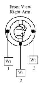
2) Subject should stand with the upper arm at the side, the elbow flexed to 90°, the forearm at a mid-position with the hand holding the ring upon which weights are attached.

3) Attach weights to the ring in one of three positions for each trial, as shown on diagram on the accompanying chart.

4) Three different weights (5, 10, and 15 lbs.), plus a reference condition of no weight will be used in each position. If 15 lbs. is too much weight to be held, smaller increments can be used. The elbow should remain flexed isometrically in each condition at 90°. No forearm rotation should be permitted with the forearm maintained in the mid-position.

5) Record the magnitude of the averaged EMGs for each load in Table 1 by analyzing the data as described above.

Questions To Ponder

1. What effect did the positions of the weights in part 3 have on the EMG results for the biceps? Plot Weight vs. EMG for each of the three torque positions and examine how biceps and brachioradialis EMG’s vary depending on the type of forearm torque applied. Knowing that the biceps muscle assists in supination, explain the results. This experiment provides data that illustrates how EMG can provides insight into how muscle recruitment is affected by the local loading conditions experienced by the body. Should tests involving EMG of elbow flexion be done in a pronated, mid, or supinated position?

Discussion

• Each of the three conditions involve no difference in the task of elbow flexion – the same weight is lifted and the joint position is the same – all that differs is the torque about the r-a joint.

• Example data for two groups and a plot of EMG vs. weight is shown above. The results are what is usually found.

• Where there is a pronatory torque applied, there is much greater activity in the biceps than with either the neutral or supinatory torque. This is an adaptation to the side action of the biceps involving supination. During elbow flexion, if the biceps is active it will also cause supination torques. With a pronatory torque it is useful to have the biceps active because the supination side action will counter the applied torque.

• For the supinatory torque, if the biceps is active some other pronatory muscle (pronator teres, quadratus, brachioradialis) will have to be active both to counter the applied supinatory torque and the side action of the biceps. Based on this EMG result, it is more efficient to shut down the biceps and let one of the other muscles do the work.

• The brachioradialis muscle EMG shows the opposite trends to the biceps EMG, being low for a pronatory torque and high for a supinatory torque. In a neutral radio-ulnar joint position, the brachioradialis will probably have little effect on pronation or supination, and the changes in activity are likely a neural compensation to what is best in terms of using the biceps. If biceps activity goes down (applied supinatory torque), some other muscle(s) has to increase flexor torque to compensate, and the brachioradialis does that. The brachialis muscle probably shows a similar pattern. Similarly, when the biceps is used at a higher level (applied pronatory torque), the brachioradialis is not needed as much and its activity decreases (probably the same for brachialis). It would be interesting to see how these activation patterns might change when the muscles get fatigued

• A major point here is that there is a sub-conscious altering of neural activation in response to the external forces applied to the joints. We don’t make a conscious decision to change how muscles are used, it just happens. Evidently there is some optimization ability within the CNS that determines the most effective pattern of muscle activation for a given situation, and we have learned this intuitively over years of developing motor skills and motor control. This can probably be generalized to any variety of movements that we might make.

What is NeuroKinetic Therapy?

NeuroKinetic Therapy is a sophisticated form of manual therapy that combines motor control theory and manual muscle testing. The science of motor control theory states that the motor control center in the cerebellum stores all the coordination patterns of the body. It is directed by the limbic system and the cerebral cortex to not only create movement patterns (such as when a baby learns to stand), but also to create substitute movement patterns when we are injured. An experiment was performed with an EMG that measured the ability of different muscles in the upper arm and forearm to perform supination of the forearm. As we know, the biceps is the most powerful supinator of the forearm. In this experiment, the biceps were inhibited and various muscles of the forearm were measured to see which one engaged to perform supination. In this case, the brachioradialis supinated the strongest. What does this tell us? When a muscle is inhibited for whatever reason, the motor control center will find a substitute muscle to perform the function. If this pattern is allowed to remain in the motor control center, dysfunction and pain will follow. How then can we undo this dysfunctional pattern?

We also know from motor control theory that if the body fails to perform a specific function, the motor control center is open to new learning for approximately 30 to 60 seconds. How then can we communicate with the motor control center? Manual muscle testing allows us to find muscles that are weak or dysfunctional in relationship to other muscles. Using the experiment above, a practitioner of NeuroKinetic Therapy would test the strength of the brachioradialis muscle and then test the strength of the biceps. In this case, the biceps would test weak. A release of 30 to 60 seconds would be performed on the brachioradialis and then the biceps would be retested. If the biceps test strong, the motor control center has been successfully reprogrammed. If the biceps still test weak, the brachioradialis could be re-released or another muscle in the area may need to be released. The client would be sent home with instructions on how to release the brachioradialis and then how to strengthen the biceps. This basic concept can be applied to muscles and dysfunctional patterns throughout the body.

NeuroKinetic Therapy is an excellent modality in rehabilitation and manual therapy because it not only identifies the cause of pain and dysfunction, but also corrects it very quickly and quite painlessly.